A Partnership Between Tumor Cells and Macrophages Drives Breast Cancer Metastasis
Cancer cells within a tumor are not uniform and display marked differences in their molecular and phenotypic characteristics, termed. . .
March 5, 2026
Introduction
Cancer cells within a tumor are not uniform and display marked differences in their molecular and phenotypic characteristics, termed intratumoral heterogeneity. These subpopulations within the tumor support tumor progression and distant metastases and are drivers of treatment resistance. In breast cancer and other cancer types, profiling these subpopulations could hold promise in designing treatment strategies to overcome this obstacle in fighting this disease1.
One phenomenon shown to expand cellular diversity is cell-cell fusion, where two cells merge into one cell. The most notable example of this is egg and sperm fusing during fertilization. Interestingly, early reports of cancer cells fusing with immune cells have been shown to promote tumor initiation, aggressiveness, and distant metastases (refer to ref. 2 for another cool study on tumor fusion cells). However, few studies have aimed to address the biological and functional characteristics of these fusion cells and clinical implications of this phenomenon. Furthermore, these fusion cells have been identified as a subpopulation in circulating tumor cells (CTCs), which are tumor cells that have left the primary tumor and entered circulation to potentially seed and outgrow at a distant metastatic site.
Goals of this study
This study builds upon a body of previous work showing that fusion cells that have been identified in circulation in cancer patients. In this study, the authors used cutting-edge technology aimed at characterizing tumors at the single cell level along with preclinical xenograft models and functional analyses to investigate these cancer fusion cells. Specific questions included:
- What are functional characteristics of these fusion cells during cancer progression and metastatic disease?
- Can this subpopulation of tumor cells be therapeutically targeted
Using single cell RNA sequencing on samples from breast primary tumors, lymph node metastases to obtain a bird’s eye view of the transcriptome of cell populations within the tumor, the authors first identified a subpopulation that expressed markers for both epithelial cells (malignant cells) and myeloid cells, prompting them to further validate this novel population. This double positive population was found in brain, liver, and lung metastases by immunohistochemistry, and in circulating tumor cells in blood from breast cancer patients. Further exploration by differential gene expression analysis showed high expression of TREM2, a common lipid-associated macrophage (LAM) marker, and using a fusion score from previous studies (references in the paper), found that the LAM signature was positively correlated with the fusion score, and this score was increased in later stages of tumor progression.
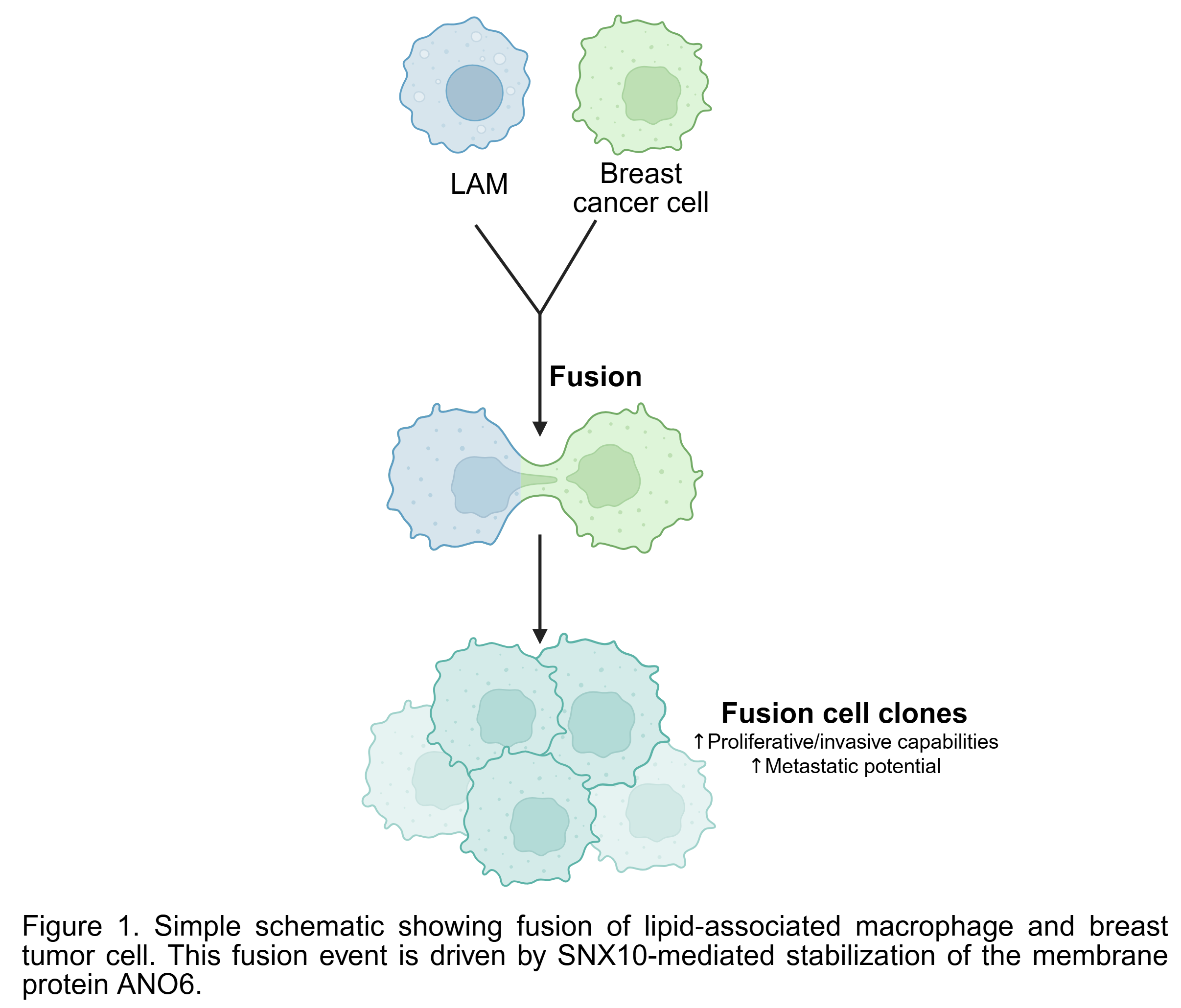
With the identification of these TREM2+ LAM-tumor fusion cells, they were next able to generate these LAM-tumor cell fusion cell an in vitro co-culture system, which allowed them to discover that these fusion cells have enhanced proliferation and invasive capabilities. They took it one step further and showed in mice that these fusion cells had enhanced metastatic potential.
Lastly, they dug deeper into the mechanism driving this fusion phenomenon, with the endosomal sorting protein known as SNX10 in mind. Identifying its interacting partner ANO6, the authors found that SNX10 enhanced stability by preventing proteasomal degradation. Using their co-culture system and knockdown strategies to show the importance of these protein-protein interactions, loss of ANO6 or SNX10 reduced fusion of LAMs and tumor cells and hence their enhanced aggressiveness in vitro.
Can these fusion cell clones be therapeutically targeted? With the idea of targeting inducers of LAMs such as oxidized LDL (oxLDL), and the evidence that statins reduce serum concentrations of oxLDL, they treated fusion cells with a stain inhibitor and found reduced aggressiveness of these fusion cells in vitro and in vivo.
This study revealed that LAMs and tumor cell fusion events drive cancer progression. Using co-culture system and in vivo models, the authors identified the axis SNX10-ANO6 on the tumor cells driving this fusion event, and it could be therapeutically targeted using an oxLDL lowering statin.
While this phenomenon was identified in breast cancer patients, it would be interesting to see the potential relevance in other cancer types. Because these fusion cells represent such a small subpopulation of clones in the tumor, they may be difficult to therapeutically target. It is also important to note that although these fusion cells increased metastatic potential, there was still a very small percentage of lung metastases in the mice. Whether the fusion event is reversible is also unknown, and because they used immunodeficient mice, how an intact immune system would affect this event is also unknown. How are these LAMs and tumor cells coming together for this event to occur? The upstream events besides the intracellular molecular mechanism identified driving fusion still require more investigation.
Article title: “Fusion of Tumor Cells with Lipid-Associated Macrophages Drives Metastatic Progression of Breast Cancer.”
Article Reference: Cheng Y, Huang G, Liu X, Wu C, Lian J, Cao J, et. al. Fusion of Tumor Cells with Lipid-Associated Macrophages Drives Metastatic Progression of Breast Cancer. Cancer Res. 2026 Feb 2;86(3):661-678. doi: 10.1158/0008-5472.CAN-25-0261. PMID: 41342370.
Additional references
Fu YC, Liang SB, Luo M, Wang XP. Intratumoral heterogeneity and drug resistance in cancer. Cancer Cell Int. 2025 Mar 18;25(1):103. doi: 10.1186/s12935-025-03734-w. PMID: 40102941; PMCID: PMC11917089.
Gast CE, Silk AD, Zarour L, Riegler L, Burkhart JG, Gustafson KT, et. al. Cell fusion potentiates tumor heterogeneity and reveals circulating hybrid cells that correlate with stage and survival. Sci Adv. 2018 Sep 12;4(9):eaat7828. doi: 10.1126/sciadv.aat7828. PMID: 30214939; PMCID: PMC6135550.
Figure created with BioRender