A crucial role of the PD-1 axis in survival and clonal selection of stem-like CD8+ T cells
For cancer cells to successfully establish and grow, they must overcome immune surveillance and immune mediated destruction. One crucial arm of the immune system, cytotoxic T lymphocytes, or CD8+ T cells, are critical players in tumor control.
January 30, 2026
Introduction
For cancer cells to successfully establish and grow, they must overcome immune surveillance and immune mediated destruction. One crucial arm of the immune system, cytotoxic T, or CD8+ T cells, are critical players in tumor control. In tumors, naïve CD8+ T cells are primed in the tumor draining lymph node (TDLN) through recognition via unique T cell receptor (TCR) expressed on their surface by tumor antigens presented to them by antigen presenting cells. Following activation, differentiated CD8+ T cells then mediate direct cancer cell killing through release of granzymes and perforin.
However, inhibitory molecules that pump the brakes on TCR signaling restrain T cell differentiation and anti-tumor function. One such signaling axis, PD-1, and its ligands PD-L1 and PD-L2, have become a target for releasing the brakes on T cells and enhancing their capacity to kill tumor cells. While immune checkpoint blockade (ICB) has shown immense success in recent years for various cancers, only a subset of patients responds and eventually relapse, highlighting a critical gap in our understanding of how PD-1 fine tunes CD8+ T cell responses.
Stem-like progenitor CD8+ T cells
While this model of CD8+ T cell priming and tumor infiltration explains some aspects of anti-tumor immunity, this model does not fully capture the diversity of T cell states within the tumor. Recent evidence has identified the presence of a subset of CD8+ T cells known as stem-like progenitor cells, which have been identified by expression of the transcription factor TCF-1, and expression of PD-1, does not fit this model, as this T cell subset resides primarily in the TDLN, have recognized antigen, and proliferated, yet do not exhibit the canonical cytotoxic function of differentiated CD8+ T cells. Instead, these stem-like CD8+ T cells serve as a pool of precursor T cells in the TDLN that can differentiate into effector T cells during ICB.
Goals of this study
This raises important questions regarding maintenance of T cell states during chronic antigen exposure. In this study published in Nature, the researchers set out to address these questions:
- What maintains these stem-like states of activated T cells in TDLNs?
- Is T cell fate imprinted early or is it being constantly shaped in the TDLNs?
The researchers used a model of subcutaneously implanted lung cancer cells expressing ovalbumin, followed by adoptive transfer of OT-1 T cells into XCR1 reporter mice. This specific model allowed the researchers to be able to mark dendritic cells through use of XCR1 and allowed them to follow tumor specific CD8+ T cells using the ovalbumin-OT-1 model. Through multiplex immunofluorescence imaging of the TDLN and flow cytometry, they found that these stem-like CD8+ T cells, marked by TCF-1, PD-1, and SLAMF6, another marker for stem-like state, were near dendritic cells, an antigen presenting cell, and restricted to the TDLN. These T cells, which were clustered with dendritic cells, had already undergone activation and proliferation, and continued to be exposed to antigen even at later stages.
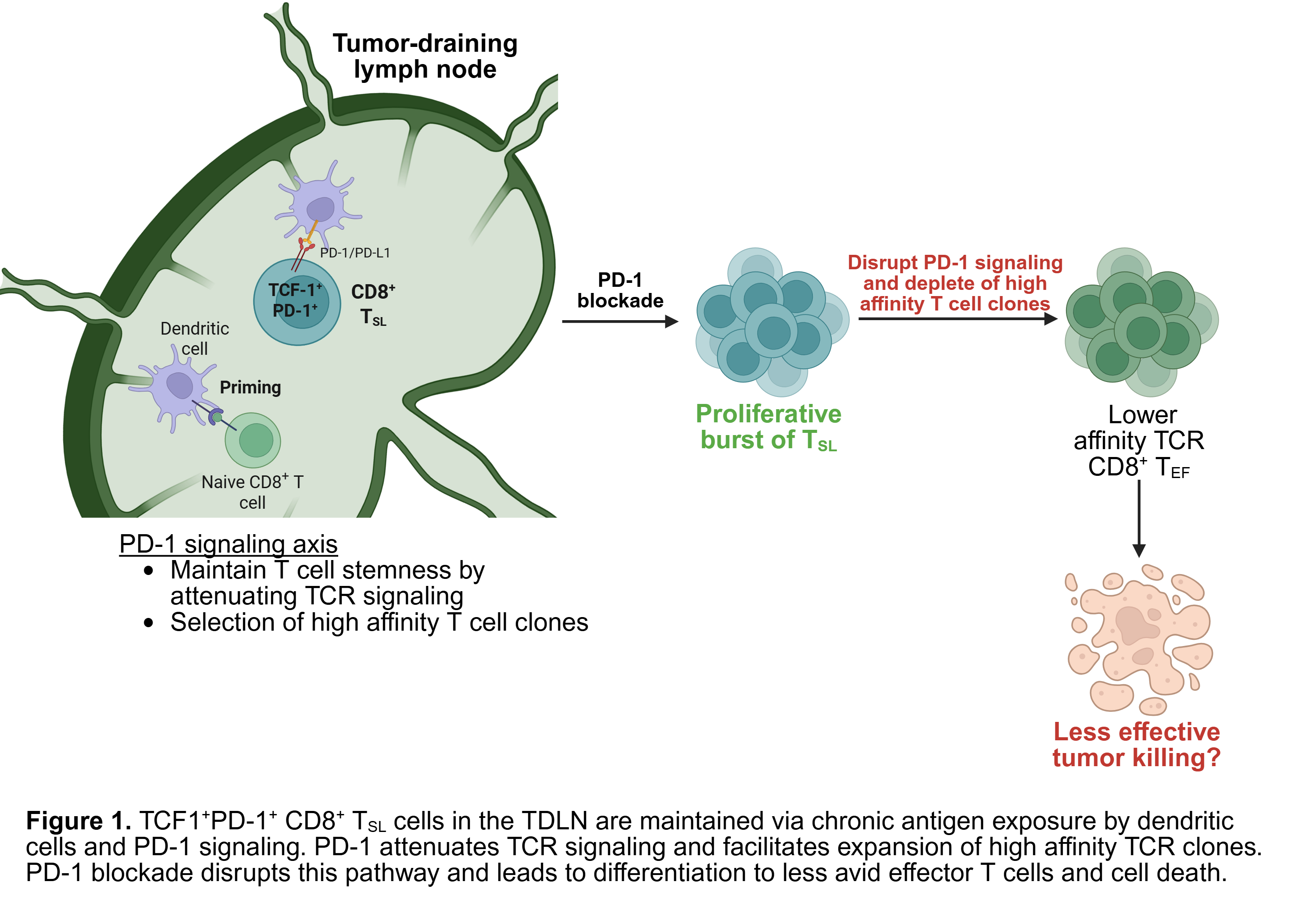
Furthermore, the authors utilized H-2Kᵇ–SIINFEKL tetramer staining to identify OVA-specific CD8⁺ T cells and generate a measure of TCR binding avidity. Interestingly, stem-like T cells with the highest TCR binding affinity also expressed the highest levels of PD-1, suggesting that PD-1 signaling may play a role in the evolution or maintenance of higher-affinity T cell clones that are better equipped to respond to tumor. Dendritic cells expressed the PD-1 ligand PD-L1, and depletion of dendritic cells reduced the stem-like CD8⁺ T cell pool. To more directly test whether blocking PD-1 depleted this population, mice were treated with PD-1 checkpoint blockade. Disruption of PD-1 signaling reduced the pool of high-affinity stem-like CD8⁺ T cell clones capable of mediating a strong anti-tumor immune response.
Overall, this study has important clinical implications for immune checkpoint blockade (ICB) therapy. Disrupting the PD-1 pathway that maintains high-affinity stem-like CD8⁺ T cells may lead to loss of this progenitor pool and potentially compromise therapeutic efficacy if lower-affinity clones instead gain access to the tumor. These findings also raise the possibility that current immunotherapies may provide short-term benefits at the cost of diminished efficacy over time due to depletion of high-affinity progenitors.
To me, this paper helps clarify the biological role of PD-1 signaling beyond its use as a marker of exhausted or dysfunctional T cells. The study convincingly demonstrates in preclinical tumor models that PD-1 signaling is protective for the maintenance of stem-like CD8⁺ T cells, and that disrupting this axis through ICB leads to depletion of higher-affinity TCR clones. However, it remains unclear whether these findings extend to other tumor types, or how these higher-affinity T cell clones overcome the immunosuppressive tumor microenvironment once they infiltrate the tumor. Moreover, this work raises the question of whether altering the timing of immunotherapy as a more cyclical treatment strategy to potentially allow time for these stem-like T cell pools to recover before another round of ICB could improve clinical outcomes.
Article title: “Inhibitory PD-1 axis maintains high-avidity stem-like CD8+ T cells.”
Article Reference: Hor, J.L., Schrom, E.C., Wong-Rolle, A. et al. Inhibitory PD-1 axis maintains high-avidity stem-like CD8+ T cells. Nature (2025). https://doi.org/10.1038/s41586-025-09440-x
Figure created in BioRender.